跳到主要內容區

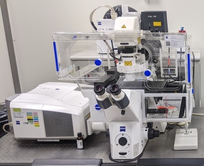
C02 掃描式雷射共軛焦顯微影像系統(Confocal Microscope System)

儀器中文全名

C02 掃描式雷射共軛焦顯微影像系統

儀器英文全名

C02 Confocal Microscope System

儀器位置

力行校區生物科技教學大樓5樓89503室

單位/教授

成大生物科技與產業科學系

陳宗嶽教授

儀器管理人

吳佳倩

TEL

06-2757575 ext. 58214-814

or ext. 58012

E-mail

okok826@hotmail.com

技術類別

£前段製程:

£微影 £鍍膜 £蝕刻 £擴散 £化學機械研磨

£表面分析:

£電子能譜儀 £表面特性

£後段製程:

£晶圓針測 £晶圓切割 £黏晶 £打線接合 £封膠

£掃描探針:

£顯微系統 £機械性質 

£形貌分析

£電子顯微:

£SEM £TEM £樣品製備

£物理性質:

£磁性 £熱分析 £電學

¢光學檢測:

£光譜儀 ¢顯微形貌分析

£生物醫學:

£分析 £樣品製備

£晶相分析:

£XRD

£其他:

£工程樣品製備 £材料力學 

£電腦計算

£分析化學:

£核磁共振儀 £質譜/層析

應用/功能

簡介

此臺顯微鏡可以得到細胞的精緻螢光影像,不同的螢光染劑發出不同的散射光,經過影像重疊的處理可以得到不同蛋白質在細胞或組織內的相關位置。

The system’s illumination and detection design allow you to simultaneously acquire multiple fluorescence signals, the relative positions of different proteins in cells or tissues can be obtained which present delicate fluorescent images.

  1. 多重螢光影像(Multi-Color Fluorescence Imaging)
  2. 多重螢光蛋白影像技術(Multi-Color GFP Imaging)
  3. 螢光共振能量轉移分析實驗(Fluorescence Resonance Energy Transfer, FRET)
  4. 長時間影像記錄(Time-Lapse Recording)
  5. 縱深掃描與測量(Z-section and measurement)
  6. 形態與動態觀察(Morphology)
  7. 3D影像擷取(3D imaging)

廠牌/型號

廠牌:Zeiss                             

型號:LSM780

重要規格

  • 倒立顯微鏡
  • 物鏡:5x、10x、20x、40x-oil、63x-oil、100x-oil
  • 激發光(nm):405、458、488、514、561、594、633
  • Detector(nm):410~750
  • 解析度:128*128、256*256、512*512、1024*1024、2048*2048、4096*4096、                               6144*6144
  • Inverted microscope
  • Objective Lens: 5x, 10x, 20x, 40x-oil, 63x-oil, 100x-oil
  • Excitation (nm): 405, 458, 488, 514, 561, 594, 633
  • Detector (nm): 410 ~ 750
  • Frame Size: 128*128, 256*256, 512*512, 1024*1024, 2048*2048, 4096*4096, 6144*6144

取樣/使用

注意事項

  • 影像系統須由陳宗嶽老師實驗室專人操作,研究人員自行準備檢體,事先預約時段,並告知實驗需求(雷射光的種類、功能)。
  • 不可使用USB,請自行攜帶光碟片存取檔案。
  • 要拍攝清晰的活細胞影像請避免使用塑膠底盤的dish或multi well plate,建議使用玻璃底盤的dish。
  • 載玻片左右兩側各保留至少0.5 cm不被蓋玻片遮蓋,以保持樣品的平整。
  • The microscope system must be operated by the operator from Prof. Tzong-Yueh Chen’s laboratory. The researcher prepares the sample, makes an appointment in advance, and informs the operator about the experimental requirements (laser, application).
  • Only CD/DVD is allowed to use for saving the data from the system computer.
  • For live-cell imaging, please avoid using plastic bottom dish or multi-well plate, the glass bottom dish is suggested.
  • The microscope slide should remain 0.5 cm on each side and not be coved by the cover glass to make the sides at the same level.

開放時段

週一至週五:09:00~12:00、13:30~16:30

操作時段以一小時為單位計算,一次預約至少一小時。

※建議先與操作員聯繫,再進行系統預約。

Monday to Friday: 9:00 ~ 12:00 and 13:30 ~ 16:30

The operation time is an hour as the unit, each reservation at least 1 hour.

※Please contact the operator before online booking

訓練課程規定

本儀器會由專人操作,不開放訓練課程

收費標準(含訓練課程)

校內學術:1500元/時

校外學術:2000元/時

業界:2000元/時

預約系統

https://cis.cfc.ncku.edu.tw/apparatus/index.php

登入成功